Introduction
Your dental X-ray tube is the beating heart of your diagnostic imaging system. Without it, there are no radiographs, no accurate diagnoses, and no treatment plans. Whether you operate a single-chair dental clinic or manage procurement for a network of dental hospitals, the performance of your dental X-ray tube directly determines the quality of patient care you can deliver.
Yet across the global dental equipment industry, tube failure remains one of the most underestimated sources of operational disruption. When a dental X-ray tube fails unexpectedly, the consequences cascade quickly: appointments are cancelled, patients are redirected, and urgent repair calls generate costs far exceeding those of a planned replacement. Industry data consistently shows that unplanned medical equipment downtime costs healthcare facilities an average of $500 to $1,000 per hour in lost productivity — and that figure does not account for the damage to patient trust or regulatory compliance risk.
The good news is that dental X-ray tube failure is rarely sudden. In most cases, the tube sends clear warning signals weeks or even months before it reaches the point of complete failure. Knowing how to recognize these signals — and acting on them promptly — is one of the most cost-effective decisions a dental practice manager, biomedical engineer, or equipment procurement specialist can make.
This guide covers the 7 most critical warning signs that your dental X-ray tube needs replacement, alongside practical diagnostic steps, maintenance best practices, and guidance on choosing the right replacement tube, including for widely used models like the CEI OPX105.
What Is a Dental X-Ray Tube and How Does It Work?
A dental X-ray tube is a vacuum-sealed glass or metal/ceramic envelope that generates ionizing radiation for diagnostic imaging purposes. Inside the tube, a heated tungsten filament (the cathode) emits a stream of electrons, which are accelerated across a high-voltage gap and directed at a tungsten or molybdenum anode target. The collision of electrons with the anode produces X-rays, which are then directed through a collimator and into the patient’s oral region to produce radiographic images.
Dental X-ray tubes fall into two primary categories:
Stationary anode tubes — the most common type used in intraoral and panoramic dental imaging. The anode is fixed in place, making these tubes simpler, more compact, and highly suited to the lower power requirements of dental applications. Our stationary anode X-ray tubes are engineered specifically for this environment.
Rotating anode tubes — used in higher-power medical imaging applications where the heat load is distributed across a spinning anode disk.
For panoramic dental imaging (OPG), the tube must rotate around the patient while continuously emitting X-rays. The panoramic dental X-ray tube is therefore subjected to unique mechanical and thermal stresses not present in standard intraoral units.
Typical Service Life
Under normal operating conditions, a dental X-ray tube has an expected service life of:
- Intraoral dental X-ray tubes: 5 to 10 years, or approximately 50,000 to 100,000 exposures
- Panoramic / OPG X-ray tubes: 3 to 7 years, depending on usage volume and maintenance practices
- High-volume clinical environments: lifespan may be significantly shorter
Factors That Affect Lifespan
Several variables influence how long a dental X-ray tube remains in reliable service:
- Daily exposure volume — high-throughput clinics accelerate wear on the filament and anode
- Warm-up protocol adherence — skipping warm-up cycles causes thermal shock to the anode
- Ambient temperature and humidity — extreme environmental conditions degrade the oil cooling medium and vacuum integrity
- Power supply stability — voltage fluctuations cause repeated stress cycles on internal components
- Maintenance frequency — irregular servicing allows minor issues to compound into critical failures
- Tube housing condition — a compromised housing allows oil leaks and radiation scatter
Understanding these factors sets the foundation for recognizing when your tube is entering its terminal phase of operation.
Warning Sign #1: Declining Image QualityWhat Causes It
Image quality degradation is the most common and clinically significant early indicator of dental X-ray tube wear. As the tungsten filament ages through repeated thermal cycles, it gradually thins and begins to evaporate, depositing tungsten molecules on the inner walls of the glass envelope. This metallic coating, known as tube “blackening,” attenuates the X-ray beam and reduces its intensity. Simultaneously, the focal spot — the precise area on the anode where electrons converge — enlarges due to filament deformation. A larger focal spot means reduced geometric sharpness in the final image.
Symptoms
- Radiographs appear progressively grainier or less sharp over weeks
- Soft tissue structures and fine bone detail become difficult to distinguish
- Images require more post-processing adjustment in the imaging software to achieve acceptable diagnostic quality
- Panoramic scans show uneven density across the image arc
- Ghost artifacts or unusual light/dark banding appears on exposures
Diagnostic Methods
- Compare recent images with archived baseline images from the same equipment taken 12 to 18 months prior
- Use a dental imaging test phantom to quantitatively assess resolution, contrast, and noise levels
- Ask the imaging software for exposure index data; a steady upward drift in required mAs values is a reliable indicator of tube output decline
- Consult your equipment’s quality assurance log if your practice maintains one (as required under radiation protection legislation in many jurisdictions)
Risks If Ignored
Persistent use of a degraded tube does not simply mean cosmetically inferior images. It means diagnostic accuracy is compromised. Missed caries, undetected periapical pathology, and inaccurate implant planning measurements can all result from poor image quality — creating both clinical and medico-legal risk.
Corrective Action
Schedule a formal image quality assessment with a qualified medical physicist or biomedical engineer. If the tube output has declined by more than 20–30% from its baseline performance, replacement planning should begin immediately.
Warning Sign #2: Increased Exposure TimeWhat Causes It
As a dental X-ray tube ages, its ability to generate sufficient X-ray output at the set exposure parameters progressively diminishes. To compensate and maintain adequate image density, operators — often unconsciously — begin increasing exposure time (mAs), tube voltage (kVp), or both. This compensatory escalation is a textbook sign of declining tube efficiency and is directly linked to filament aging and anode surface pitting.
Symptoms
- Technicians or dentists regularly increase exposure settings to achieve the same image quality
- The automatic exposure control (AEC) system on modern panoramic units repeatedly selects maximum or near-maximum exposure values
- Exposure times that were once 60–70ms for a standard periapical view have crept to 90–110ms or beyond
- Patients receive higher radiation doses than the equipment’s published specifications indicate
Diagnostic Methods
- Maintain a logbook of exposure parameters for each imaging modality. A consistent upward trend in required exposure values over a 3–6 month period is a definitive diagnostic signal.
- Compare current kVp and mAs settings against the manufacturer’s recommended baseline exposure charts for your specific unit
- For panoramic units, review the AEC selection history in the system logs if available
Risks If Ignored
Increasing exposure time directly translates to increased patient radiation dose. This conflicts with the ALARA (As Low As Reasonably Achievable) principle that governs radiation protection in dental practice worldwide. Regulatory inspections that identify unjustifiably elevated patient doses can result in equipment suspension and compliance notices.
Corrective Action
Document the exposure escalation trend and present it to your equipment service provider. Cross-reference with image quality data. In most cases, if both image quality and output efficiency have declined simultaneously, tube replacement is the appropriate course of action.
Warning Sign #3: Frequent Equipment Error MessagesWhat Causes It
Modern dental panoramic and CBCT units are equipped with sophisticated self-monitoring systems that track tube parameters including filament current, anode voltage, tube temperature, and exposure cycle counts. As internal tube components degrade, these monitoring systems begin generating error codes — initially intermittent, but increasing in frequency as the tube approaches end of life.
Symptoms
- The imaging console displays recurring “tube warm-up failure” or “exposure aborted” error messages
- Error codes appear even after the prescribed warm-up cycle has been completed
- The system requires multiple attempts before successfully completing an exposure
- The unit enters protective shutdown mode during patient positioning
- Error logs show a pattern of escalating fault frequency over a 30–90 day period
Diagnostic Methods
- Export and review the equipment error log. Most major OPG manufacturers (Planmeca, Vatech, Carestream, Sirona/Dentsply) provide service-level log access for authorized engineers.
- Note whether error codes are tube-specific (filament, anode, HV generator) or system-wide. Tube-specific errors that cannot be resolved by recalibration or software reset indicate hardware degradation.
- Contact your equipment service partner with the specific error codes for accurate diagnosis
Risks If Ignored
Operating equipment that is generating repeated error messages introduces unpredictability into the clinical workflow. An unexpected shutdown during a patient exposure — particularly during a CBCT scan — may require repeat imaging, doubling the patient’s radiation dose. Equipment that errors repeatedly may also be operating outside its safe parameters, creating potential radiation safety risks.
Corrective Action
Do not disable or override error monitoring systems. Treat recurring tube-specific error codes as a formal indication to begin the tube replacement process.
Warning Sign #4: Overheating During OperationWhat Causes It
Every dental X-ray tube generates heat as a byproduct of X-ray production — typically around 99% of the electrical energy input converts to heat rather than X-rays. Under normal conditions, this heat is managed through the tube’s oil-cooling system and the thermal mass of the anode. As the tube ages, three failure modes can cause overheating: oil degradation (reducing its cooling capacity), vacuum deterioration (allowing trace gases that transfer heat abnormally), and anode pitting (creating hot spots on the focal track).
Symptoms
- The tube housing feels unusually hot to the touch after a standard examination sequence
- The equipment console displays “tube overtemperature” or “thermal limit” warnings
- The system enforces mandatory cooling delays between exposures that were not previously required
- Oil leakage is visible around the tube housing seals — a serious indicator of housing integrity failure
- The ambient temperature around the X-ray unit rises noticeably during a normal clinical session
Diagnostic Methods
- Use a non-contact infrared thermometer to monitor tube housing surface temperature during and after typical exposure sequences. Compare readings against the manufacturer’s specifications.
- Inspect the housing for oil residue around cable entry points and the collimator interface
- Check whether mandated cooling times between exposures have increased compared to when the unit was new
- A qualified engineer can measure the tube’s actual duty cycle and compare it against design specifications
Risks If Ignored
Chronic overheating accelerates every other failure mode simultaneously. It degrades the dielectric oil faster, contributes to vacuum deterioration, and can cause the glass envelope to crack — resulting in complete and irreversible tube failure. In worst-case scenarios, a cracked tube envelope can cause electrical arcing within the housing.
Corrective Action
If oil leakage is identified, the tube should be taken out of service immediately. Overheating without visible leakage still warrants urgent engineer assessment. Do not continue operating an overheating tube by simply extending cooling intervals — this treats the symptom rather than the cause.
Warning Sign #5: Unusual Noises or Electrical IssuesWhat Causes It
A functioning dental X-ray tube operates silently or with minimal noise. Unusual sounds during operation indicate mechanical or electrical anomalies within the tube or its associated high-voltage circuitry. The most significant of these is electrical arcing — a high-frequency crackling or snapping sound produced when residual gas molecules inside the tube allow electrons to ionize the gas and create uncontrolled electrical discharges.
Symptoms
- An audible crackling, snapping, or popping sound during exposures
- A visible flash or flicker at the tube housing during operation (observable in a darkened room)
- Circuit breakers or fuses in the X-ray generator tripping repeatedly
- Intermittent or flickering imaging on the sensor or film before complete exposure failure
- A burning or ozone smell in the vicinity of the tube or generator
- Sparking at the high-voltage cable connections
Diagnostic Methods
- Operate the unit in a semi-darkened environment with an engineer present to visually inspect for arcing
- Review the generator’s fault log for high-voltage trip events
- Inspect high-voltage cables and receptacles for signs of tracking (carbon deposit trails indicating previous arcing). Our 75KVDC high-voltage cables are designed to withstand these stresses, but they too must be inspected regularly as part of any comprehensive tube assessment.
- An engineer can perform insulation resistance testing on the HV cable and receptacle assembly to isolate whether arcing originates in the tube or the cable
Risks If Ignored
Arcing represents an imminent catastrophic failure risk. An uncontrolled electrical discharge can destroy the X-ray generator, damage the imaging detector, and potentially create a fire hazard. Equipment exhibiting active arcing should be taken out of service immediately and not operated until a full assessment has been completed.
Corrective Action
Do not attempt to continue using equipment that produces audible arcing sounds. Isolate the unit, document the fault, and contact a qualified service engineer immediately.
Warning Sign #6: Inconsistent Radiation OutputWhat Causes It
Radiation output consistency is fundamental to diagnostic reliability. A well-functioning dental X-ray tube delivers a reproducible, stable beam at the same output level for every exposure at identical settings. As the filament degrades and the anode surface becomes pitted, output variability increases — a phenomenon clinically described as “beam instability.” This can also result from aging of the high-voltage generator components, but in many cases the tube itself is the primary source.
Symptoms
- Repeated exposures at identical settings produce images of noticeably different density
- Sensitometry readings (using a step wedge on film or digital systems) show high variability between consecutive exposures
- The imaging software’s exposure index varies significantly between identical views taken on the same day
- Some exposures are significantly overexposed while others are underexposed, despite no change in technique factors
Diagnostic Methods
- Perform a reproducibility test: take 10 consecutive exposures at identical kVp, mAs, and geometry settings using a calibrated dosimeter. Calculate the coefficient of variation (CV) of the output measurements. A CV greater than 5% indicates clinically significant instability.
- Compare dosimeter readings against the unit’s published output specifications
- If the generator parameters are stable but output remains variable, the tube is the likely source
Risks If Ignored
Inconsistent output means diagnostic reliability is unpredictable on a shot-by-shot basis. Patients may receive unnecessarily high doses during overexposed shots. Underexposed images may require retakes, further increasing cumulative patient dose. From a regulatory standpoint, output inconsistency is a calibration failure that may trigger enforcement action during radiation protection inspections.
Corrective Action
Formal dosimetric testing by a medical physicist is recommended. If output inconsistency is confirmed and cannot be resolved through generator calibration, tube replacement is indicated.
Warning Sign #7: Rising Maintenance and Repair CostsWhat Causes It
From a financial management perspective, the total cost of ownership of any X-ray tube follows a predictable bathtub curve. Costs are relatively low through the tube’s productive midlife, but rise sharply as the tube enters its wear-out phase. Repeated service calls for the same recurring issues — particularly those involving tube-related faults — are a clear economic signal that the tube has reached the end of its cost-effective service life.
Symptoms
- The unit has required 3 or more unplanned service visits in the past 12 months for tube or imaging-related faults
- Repair invoices reference recurring issues such as filament calibration, HV arcing, or output instability
- Parts costs are increasing because the tube model is aging and spares are becoming harder to source
- Each repair provides only a short period of reliable operation before the next fault occurs
- The total cost of the last 2–3 repair events approaches or exceeds the cost of a replacement tube
Diagnostic Methods
- Compile a 24-month maintenance cost history for the specific unit. Separate tube-related costs from unrelated mechanical or software issues.
- Calculate the repair-to-replacement cost ratio: if cumulative repair costs over 18–24 months exceed 60–70% of a replacement tube’s cost, replacement is the financially rational choice.
- Request a written technical assessment from your service engineer documenting the root cause of recurring faults
Risks If Ignored
Continuing to invest in a failing tube is not simply a question of economics. Each repair buys a progressively shorter window of reliable operation, and the probability of unexpected catastrophic failure — with all the associated clinical disruption — increases with each repair cycle. The risk of a complete failure during a critical patient examination, with no replacement available, creates both clinical and reputational risk.
Corrective Action
Engage a specialist dental X-ray tube supplier to obtain a formal replacement recommendation and cost comparison. Proactive replacement planning allows you to schedule the changeover during a low-volume clinical period, avoiding the disruption of emergency replacement.
Repair vs. Replace: Which Option Makes More Sense?
The decision to repair or replace a failing dental X-ray tube requires careful analysis across multiple dimensions. The following comparison provides a structured framework for this decision.
| Factor | Repair | Replace |
|---|---|---|
| Upfront Cost | Lower | Higher (full tube cost) |
| Downtime | Variable; parts availability can extend delays | Predictable; planned installation typically 1–2 days |
| Reliability Post-Intervention | Moderate; often temporary; root cause may persist | High; full performance restoration from day one |
| Safety | Risk remains if underlying degradation continues | Risk fully reset; full radiation safety compliance |
| Warranty | Typically no warranty on repaired components | New tube warranty (typically 6–12 months) |
| Long-term ROI | Poor if repair is the third or more event | Strong; eliminates escalating repair cycle |
| Image Quality | Partial improvement at best | Full restoration to manufacturer specification |
| Regulatory Compliance | May still fail dosimetric audit | Fully compliant from point of installation |
Verdict: If a tube has required more than two significant repairs, or if cumulative repair costs over 24 months have exceeded 50% of replacement cost, replacement is the financially and clinically superior choice in virtually all cases.
How to Extend the Life of Your Dental X-Ray Tube
Proactive maintenance is the single most effective strategy for maximizing your dental X-ray tube’s service life. The following best practices are recommended by equipment manufacturers and supported by decades of field experience.
Daily Maintenance Tips
- Inspect the tube housing visually before the first use each day for any signs of oil leakage, physical damage, or cable wear
- Ensure the collimator opening is clean and unobstructed
- Confirm that the unit’s cooling fan (where fitted) is operational
- Log any unusual noises, error messages, or image quality changes at the end of each clinical day
Proper Warm-Up Procedures
Warm-up is one of the most important — and most commonly neglected — aspects of dental X-ray tube care. Thermal shock from cold starts is a leading cause of premature filament failure.
- Follow the manufacturer’s prescribed warm-up protocol every morning before the first patient exposure
- Begin with low-kVp, low-mAs exposures and step up progressively
- Never perform high-exposure panoramic or CBCT scans immediately after system startup
- If the system has been idle for more than 4 hours, treat it as a cold start and run the full warm-up sequence
Environmental Controls
- Maintain the X-ray room temperature between 18°C and 24°C (64°F–75°F); high ambient temperatures reduce the cooling differential and accelerate tube wear
- Keep relative humidity below 70% to protect electronic components and prevent condensation on the tube housing
- Protect the unit from direct sunlight, which can raise surface temperatures and cause UV degradation of rubber cable insulation
- Ensure adequate ventilation around the tube housing; do not allow storage items to obstruct airflow
Usage Best Practices
- Never exceed the tube’s rated duty cycle; allow mandatory cooling periods between high-load exposure sequences
- Use the minimum kVp and mAs settings that produce diagnostically adequate images (ALARA principle)
- Avoid mechanical shock to the tube housing; panoramic units are particularly vulnerable when the rotating arm is moved carelessly
- Train all clinical staff in proper equipment handling and emergency shutdown procedures
Preventive Maintenance Schedule
| Frequency | Action |
|---|---|
| Daily | Visual inspection, warm-up protocol, error log review |
| Monthly | Cable and connector inspection, housing surface cleaning |
| Quarterly | Dosimetric output verification, image quality phantom test |
| Annually | Full engineer inspection, kVp and timer calibration, HV cable insulation test, oil level check (where applicable) |
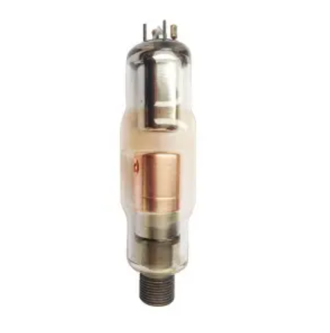
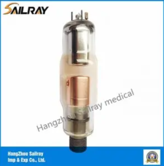
When Should You Replace a CEI OPX105 Dental X-Ray Tube?
The CEI OPX105 is a widely used stationary anode X-ray tube designed for panoramic dental imaging systems. It has established itself as a reliable workhorse in OPG units across Europe, Asia, and the Middle East, and is used by numerous equipment OEMs and independent service organizations.
Performance Indicators Specific to the OPX105
Under typical clinical conditions (20–40 panoramic exposures per day), a CEI OPX105 tube typically delivers:
- Expected service life: 4 to 6 years
- Approximate exposure count at end of life: 60,000 to 90,000 panoramic cycles
- Output degradation threshold requiring action: ≥25% decline from commissioning output
Common Failure Modes
Field data from service organizations indicates that CEI OPX105 tubes most commonly fail through the following mechanisms:
- Filament burn-out — the most frequent failure mode; often preceded by gradual output reduction and increased exposure time requirements
- Glass envelope blackening — occurs in tubes exceeding 70,000 exposures; produces the characteristic image quality degradation described in Warning Sign #1
- Anode surface pitting — accelerated in units where warm-up protocols are not consistently followed; produces output variability (Warning Sign #6)
- HV insulation breakdown — associated with units operating in high-humidity environments or with aged dielectric oil
Replacement Recommendations
Replace a CEI OPX105 tube when any of the following conditions are met:
- Tube has exceeded 5 years of service in a high-volume practice (30+ panoramic exposures/day)
- Two or more of the 7 warning signs described in this guide are simultaneously present
- Dosimetric testing confirms output has declined by 25% or more from baseline
- The unit has required 2 or more tube-related service interventions in a 12-month period
- The existing tube model is approaching obsolescence and spare availability is declining
For OEM manufacturers and equipment distributors seeking compatible replacement solutions, our range of panoramic dental X-ray tubes includes high-quality alternatives to the CEI OPX105, manufactured to the same dimensional and electrical specifications required for drop-in compatibility.
FAQ
Q1: How long does a dental X-ray tube last?
A: Most dental X-ray tubes have a service life of 5 to 10 years for intraoral units and 3 to 7 years for panoramic (OPG) tubes under normal clinical usage. High-volume practices will typically see shorter lifespans due to greater daily exposure counts and thermal cycling. Proper warm-up procedures and adherence to preventive maintenance schedules can extend service life significantly.
Q2: Can a dental X-ray tube be repaired?
A: Minor issues such as filament recalibration or high-voltage cable replacement can sometimes extend tube life. However, the tube envelope itself — the sealed vacuum assembly — cannot be meaningfully repaired once it has degraded internally. In most cases where the tube has experienced filament burn-out, glass blackening, or anode pitting, replacement is the only reliable solution. Repeated repairs of the same tube typically indicate it has reached end of life.
Q3: What causes X-ray tube failure?
A: The primary causes of dental X-ray tube failure are filament aging (from repeated thermal cycles), anode surface pitting (from inadequate warm-up and high-load cycling), dielectric oil degradation (which reduces cooling efficiency), and vacuum deterioration (which allows internal arcing). Environmental factors such as high ambient temperature, humidity, and unstable power supply accelerate all of these mechanisms.
Q4: How often should dental imaging equipment be inspected?
A: A formal engineer inspection should be performed at least annually, including kVp and timer calibration, dosimetric output verification, and HV cable testing. Quarterly dosimetric output checks using a calibrated dosimeter are recommended for high-volume practices. Daily visual inspection and warm-up logging should be standard practice in every clinical environment.
Q5: What are the risks of using an aging X-ray tube?
A: An aging dental X-ray tube poses three categories of risk: clinical (reduced image quality leading to diagnostic error), safety (increased patient radiation dose due to output inconsistency and exposure escalation), and operational (unexpected equipment failure causing unplanned downtime). Regulatory risk is also significant — radiation protection legislation in most countries requires that imaging equipment operate within defined performance parameters, and a degraded tube that fails dosimetric audit can result in equipment suspension.
Q6: How do I know if my panoramic X-ray tube specifically needs replacing?
A: Panoramic tubes show early failure signs through image arc banding , increased motor positioning errors, and AEC system selecting maximum exposure values. Because panoramic tubes rotate during exposure, mechanical wear is also a factor — listen for bearing noise from the rotating arm. Any combination of image quality decline and exposure escalation in a panoramic unit is a strong indication that tube replacement assessment is needed.
Q7: What is the difference between a stationary anode and a rotating anode X-ray tube in dental applications?
A: Stationary anode tubes are used in the vast majority of dental applications — both intraoral and panoramic — because dental imaging requires relatively low power levels. The anode remains fixed, making the tube simpler, more compact, and cost-effective. Rotating anode tubes, where the anode disk spins to distribute heat across a larger surface, are primarily used in higher-power medical imaging modalities such as CT. Dental OPG systems exclusively use stationary anode designs.
Q8: Can I replace a dental X-ray tube myself?
A: No. Dental X-ray tube replacement involves disconnection of high-voltage cables , handling of radiation-producing equipment, and subsequent dosimetric calibration verification. This work must be performed by a qualified biomedical engineer or authorized equipment service technician. In most jurisdictions, radiation-producing equipment may only be serviced by licensed personnel, and a post-replacement radiation safety survey is legally required before returning the unit to clinical use.
Q9: How much does dental X-ray tube replacement cost?
A: Replacement tube costs vary significantly depending on tube type, manufacturer, and supply source. Panoramic OPG replacement tubes from OEM suppliers typically range from $800 to $3,000 USD, while aftermarket-compatible replacements from qualified manufacturers can offer equivalent performance at 30–50% lower cost. Total replacement costs including engineer installation and dosimetric recalibration typically range from $1,200 to $5,000 depending on the unit model and geographic location.
Q10: Where can I source a reliable replacement dental X-ray tube?
A: Replacement tubes can be sourced directly from the original equipment manufacturer (OEM), from specialist dental X-ray component suppliers, or from authorized distributors. For international procurement, it is important to verify that the replacement tube meets the dimensional, electrical, and radiation output specifications of the original. Suppliers should be able to provide technical datasheets confirming compatibility and should offer post-sales technical support. Explore our full dental X-ray tube product range for OEM-compatible replacement solutions across a wide range of panoramic and intraoral dental imaging systems.
Conclusion
The dental X-ray tube is one of the most critical — and most frequently overlooked — components in a dental practice’s diagnostic infrastructure. The 7 warning signs detailed in this guide — declining image quality, increased exposure time, frequent error messages, overheating, unusual noises, inconsistent radiation output, and escalating repair costs — collectively provide a reliable early warning system that any clinician, biomedical engineer, or procurement manager can use to make timely, evidence-based replacement decisions.
Early action is always more cost-effective than emergency response. A planned tube replacement, budgeted and scheduled in advance, costs a fraction of the combined disruption, emergency service fees, and patient impact associated with unplanned tube failure. It also ensures continuous radiation safety compliance — a non-negotiable requirement in every dental practice jurisdiction worldwide.
Assess your current dental X-ray equipment honestly against the criteria in this guide. If you recognize two or more of the warning signs described above, do not delay — schedule a formal inspection with a qualified biomedical engineer or contact a specialist supplier to discuss your replacement options.
For dental equipment distributors, OEM manufacturers, and procurement managers sourcing high-quality replacement dental X-ray tubes with reliable technical support, we invite you to contact our team directly. Our specialists can assist with compatibility verification, technical documentation, and supply chain solutions tailored to your specific equipment and volume requirements.
Media Contact
Company Name: Sailray Medical System Co., Ltd.
Email: Send Email
Country: China
Website: https://www.dentalx-raytube.com/